- 30 May 2022
- Posted by: nemcatgroup
- Category: Publications

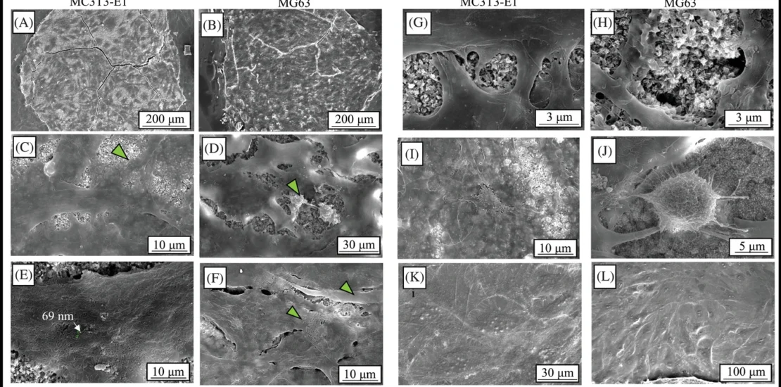
A contiguous carbonate-rich hydroxyapatite microcoating in a microfluidic device represents a substrate that has chemical and structural similarity to bone mineral. The present work describes a low-temperature method to deposit a carbonate-rich hydroxyapatite microcoating on a glass slide and its incorporation within the microchannels of a microfluidic device. A glass slide is covered/masked with polypropylene-based tape and CaCO3 nanoparticles are deposited on exposed areas by convective self assembly. The precursor CaCO3 is converted to carbonate-rich hydroxyapatite by dissolution-recrystallization in phosphate-buffered saline. The microcoating is aligned/incorporated within a microchannel when the underlying glass is bonded to a polydimethylsiloxane structure with the device layout. X-ray diffraction, laser Raman microspectroscopy, and X-ray photoelectron spectroscopy indicate that the microcoating was comprised of carbonate-rich hydroxyapatite. Scanning electron microscopy and 3D laser confocal microscopy showed that was comprised of nanocrystalline rod-like clusters that collectively exhibit a thickness of ∼20 µm. Monocultures/cocultures of osteoblast-lineage (MC3T3-E1, MG63) and preosteoclast-lineage (RAW 264.7) cells were performed. Osteoblast-lineage cells adhered to the microcoating and deposited an extracellular matrix of collagen fibrils and mineral accretions. Mineralization was detected in/near the inlet wells. The microcoating is analogous to bone mineral and could be applied to various layouts and mineral systems.
